Home
/ Leg Muscle Diagram / 13 700 Best Leg Muscle Anatomy Images Stock Photos Vectors Adobe Stock / Legs are used for standing, and all forms of.
Leg Muscle Diagram / 13 700 Best Leg Muscle Anatomy Images Stock Photos Vectors Adobe Stock / Legs are used for standing, and all forms of.
Leg Muscle Diagram / 13 700 Best Leg Muscle Anatomy Images Stock Photos Vectors Adobe Stock / Legs are used for standing, and all forms of.. This important tendon in the back of the calf and ankle stores the elastic energy needed for running, jumping, and other physical activity. The short head originates from the lateral lip of linea aspera and. You may also find transversus abdominis, iliopsoas, gluteus medius, pectineus, adductor longus. Causes of upper leg pain related to trauma may include the following. The aim of this exercise is to improve your confidence in identifying different structures.
It is also visible on the medial edge of the thigh from the anterior. The largest muscle masses in the leg are present in the thigh and the calf. Your leg muscles are some of the hardest working muscles in your body. Observe the leg muscle diagram posted above and notice that there are many parts in the muscles. One of the most important tendons in terms of mobility of the leg is the achilles tendon.
Human Leg Muscles Diagram Koibana Info from i.pinimg.com The 3 muscles are called triceps coxae. Flexes elbow and moves forearm. The human leg, in the general word sense, is the entire lower limb of the human body, including the foot, thigh and even the hip or gluteal region. Brings leg back to and across body. The aim of this exercise is to improve your confidence in identifying different structures. Your leg muscles are some of the hardest working muscles in your body. Brings hip away from body. Legs are used for standing, and all forms of.
Mccarthy, ph.d., professor of physical therapy at the university of alabama at birmingham.
Like the quadriceps, the hamstring muscle group also contains four separate muscles: It acts as a tensor of the arches of the foot, but can also be added with the first digit and plantar flexion of its first phalanx. Your leg muscles are some of the hardest working muscles in your body. Leg muscles diagram labeled : Flexes elbow and moves forearm. There are many muscles located in the lower leg, but there are three that are particularly well known—the gastrocnemius and the soleus, which are the most powerful muscles in the lower leg, and the anterior tibialis. In the leg muscles diagram above, there are many muscles that make up your legs and support it to move. The gastrocnemius is the larger calf muscle, forming the bulge visible beneath the skin. The muscles that make up the quadriceps are the strongest and leanest of all muscles in the body. Raises and rotates arm in all directions. From the large, strong muscles of the buttocks and legs to the tiny, fine muscles of the feet and toes, these muscles can exert tremendous power while constantly making small adjustments for balance — whether. The groin muscles are a group of muscles situated high on the leg in the inner thigh. These four muscles at the front of the thigh are the major extensors (help to extend the leg.
The gastrocnemius is the larger calf muscle, forming the bulge visible beneath the skin. Take a look at the leg muscles diagram below, where you see each muscle clearly labeled. Raises and rotates arm in all directions. One of the most important tendons in terms of mobility of the leg is the achilles tendon. Observe the leg muscle diagram posted above and notice that there are many parts in the muscles.
Leg Muscle Anatomy Function Facts Openfit from cdn.prod.openfit.com There are many muscles located in the lower leg, but there are three that are particularly well known—the gastrocnemius and the soleus, which are the most powerful muscles in the lower leg, and the anterior tibialis. It acts as a tensor of the arches of the foot, but can also be added with the first digit and plantar flexion of its first phalanx. On the medial edge of the posterior thigh is the gracilis muscle. These muscles start at the bottom of your pelvis extending down the back of your thigh and along either side of your knee, to your lower leg bones. Extends spine and trunk back. Pain in your calf or thigh can be caused by muscle cramps, a pulled or strained muscle, or issues related to your nerves. You may also find transversus abdominis, iliopsoas, gluteus medius, pectineus, adductor longus. For images of the muscle, click on each link under location.
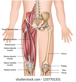
In this image, you will find rectus abdominis, external oblique, inguinal ligament, tensor fascia lata, gracilis, sartorius, rectus femoris, the iliotibial band in it.
The gastrocnemius is the larger calf muscle, forming the bulge visible beneath the skin. This is why you have to indicate which biceps you are taking about when discussing one or other of these muscles. From the large, strong muscles of the buttocks and legs to the tiny, fine muscles of the feet and toes, these muscles can exert tremendous power while constantly making small adjustments for balance — whether. Calf muscle anatomy, calf muscle picture anatomy, lateral calf muscles, lower leg muscle anatomy, medial calf muscles, what are the two calf muscles called, what are your calf muscles called, where is your calf muscle, human muscles, calf muscle anatomy, calf muscle picture anatomy, lateral calf. The 3 muscles are called triceps coxae. You may also find transversus abdominis, iliopsoas, gluteus medius, pectineus, adductor longus. See more ideas about muscle anatomy, human anatomy and physiology, body anatomy. Pain in your calf or thigh can be caused by muscle cramps, a pulled or strained muscle, or issues related to your nerves. Related posts of muscles and tendons of the leg elbow muscle anatomy mri. Collectively referred to as the hip adductors, the groin muscles are responsible for adduction of the hip, or drawing the leg in. Leg muscles diagram labeled : On the medial edge of the posterior thigh is the gracilis muscle. The hamstring muscles, also known as the rear thighs, make up the backside of the upper leg anatomy.
Muscle of the human leg diagram in this image, you will find muscle of the human leg diagram, hip and femur middle layer, hip and femur deep layer, overview of the most important muscles of the leg, femur middle layer, femur deep layer, rectus femoris m. Elbow muscle anatomy mri 12 photos of the elbow muscle anatomy mri elbow muscle anatomy axial, elbow muscle anatomy mri, human muscles, elbow muscle anatomy axial, elbow muscle anatomy mri For women, shaping the thigh muscles is an essential goal of physical fitness. Extends spine and trunk back. See more ideas about muscle anatomy, human anatomy and physiology, body anatomy.
Quadriceps Muscle Images Stock Photos Vectors Shutterstock from image.shutterstock.com Calf muscle anatomy, calf muscle picture anatomy, lateral calf muscles, lower leg muscle anatomy, medial calf muscles, what are the two calf muscles called, what are your calf muscles called, where is your calf muscle, human muscles, calf muscle anatomy, calf muscle picture anatomy, lateral calf. These four muscles at the front of the thigh are the major extensors (help to extend the leg. Legs are used for standing, and all forms of. The calf muscle, on the back of the lower leg, is actually made up of two muscles: Leg muscles diagram labeled : The largest muscle masses in the leg are present in the thigh and the calf. Extends spine and trunk back. This is why you have to indicate which biceps you are taking about when discussing one or other of these muscles.
The achilles tendon is also located in the lower leg.
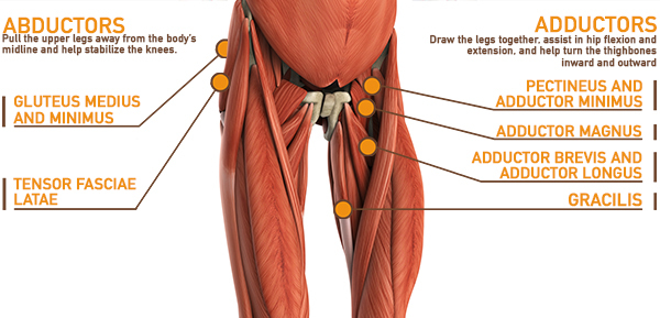
The biceps femoris is a muscle of the posterior thigh composed of a long head and a short head. This is why you have to indicate which biceps you are taking about when discussing one or other of these muscles. It acts as a tensor of the arches of the foot, but can also be added with the first digit and plantar flexion of its first phalanx. Leg muscles diagram labeled : Flexes elbow and moves forearm. Pain in your calf or thigh can be caused by muscle cramps, a pulled or strained muscle, or issues related to your nerves. Diagram illustrating muscle groups on back of human legs. The adductors work virtually any time your legs are active, whether for standing, squatting, lunging, and most other leg moves. Biceps femoris (long head) biceps femoris (short head) semitendinosus. It is also visible on the medial edge of the thigh from the anterior. These muscles start at the bottom of your pelvis extending down the back of your thigh and along either side of your knee, to your lower leg bones. This group includes the adductor magnus, adductor longus, and adductor brevis muscles, as well as the pectineus and gracilis. On the medial edge of the posterior thigh is the gracilis muscle.
